
Hysteroscope is a telescope that allows a gynecologist to look inside your uterus. The hysteroscope is a long thin tube, which has a built-in viewing device. Hysteroscopy is useful for diagnosing and treatment of endometrial polyp, submucus fibroids, menstrual bleeding, endometrial adhesions, endometrial celluar abnormalities, premalignant and malignant conditions.
Hysteroscopy can help treating some problems that cause infertility, miscarriages, and abnormal menstrual bleeding.
The best view of the uterine lining is obtained during the week that follows your period. If you have regular cycles and are able to anticipate the timing of your next period you can plan to have the hysteroscopy done in the following week.
How is Hysteroscopy done?
You lie on your back on an examination / operating table, with your knees bent and supported or your feet in footrests, as you would for a pelvic examination. Your vagina is cleaned with an antibacterial lotion.
You may require local, regional, or general anesthesia.
Local anesthesia: Injections of numbing medicine is given into the tissue surrounding your cervix, deep in your vagina. You remain awake through the procedure. You may feel pelvic cramping as period pain.
Regional anesthesia: Either with a spinal or an epidural block. An anaesthetic is injected in your lower back by an anaesthetist. You are awake for the procedure, but you do not feel pain from your pelvic region.
General anesthesia: Is required if you are having other procedures such as laparoscopy done at the same time as hysteroscopy. You will be sleep so you are unconscious during the procedure. General anesthesia is administered by an anaesthetist, who gives you intravenous drugs followed by breathing a mixture of gases through a mask. After the anesthetic takes effect, a tube may be put down your throat to help you breathe and administer the anaesthetic drug.
The cervical os (opening of your cervix) may need to be dilated or stretched to widen it. The hysteroscope is inserted through the vagina and cervix and into the uterus. A liquid or gas is released through the hysteroscope to expand the inside of the uterus so that your doctor has a clearer view. A light at the end of the instrument allows your doctor to see the walls of the uterus and the openings of the fallopian tubes at the top of the uterus.
Your gynaecologist may also insert small surgical instruments through the hysteroscope to carry out such procedures as taking a sample of tissue (biopsy), removing a polyp or growth or IUD, dividing adhesions, removing the lining of the uterus with dilation and curettage (D&C), or treating the lining of your uterus with ablation to reduce or prevent bleeding.

|
|
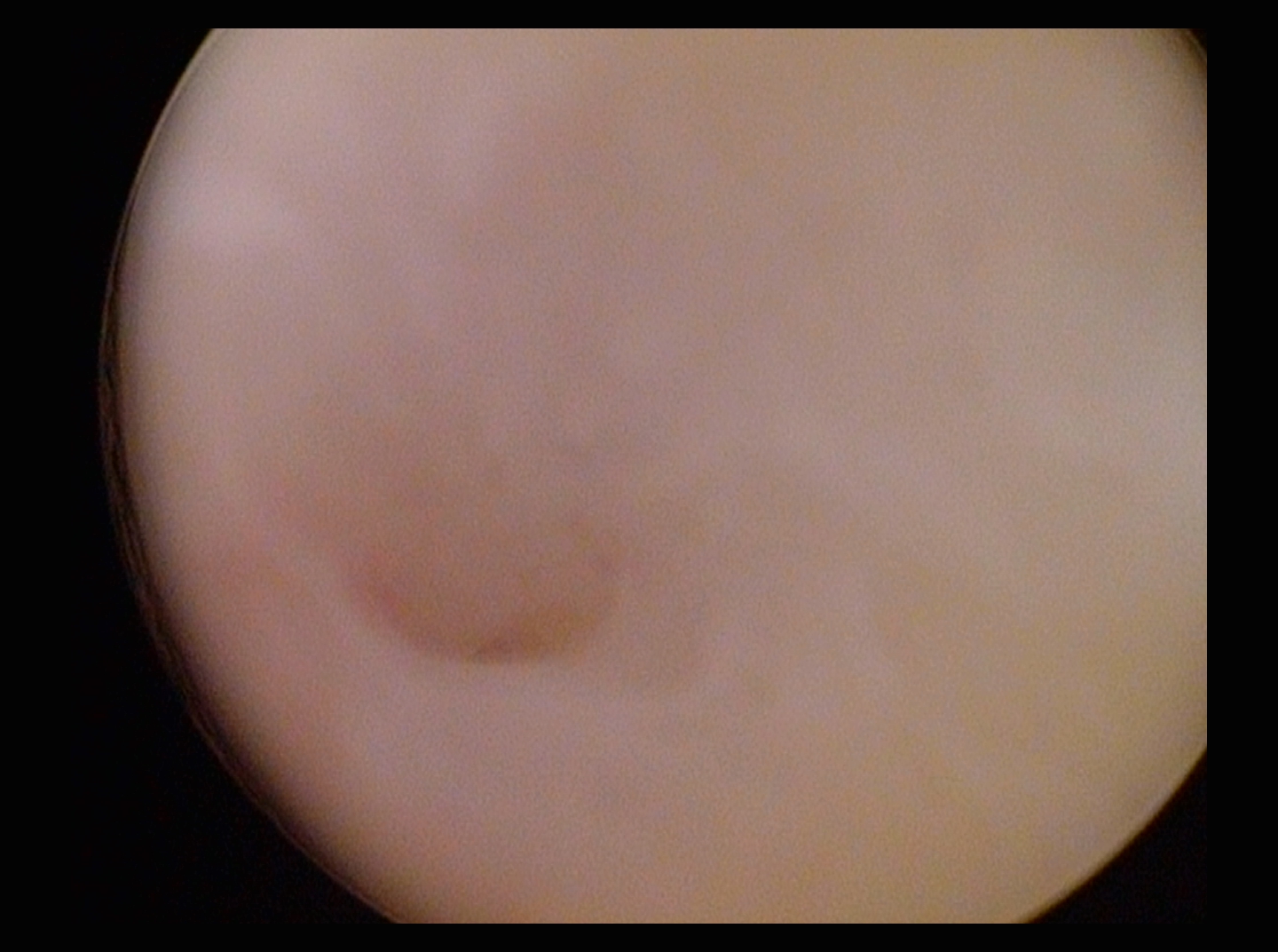
Hysteroscopy Normal Fallopian Tube Ostium Left
|
|
|
|
Rt Tube Ostium
|
|
|
|
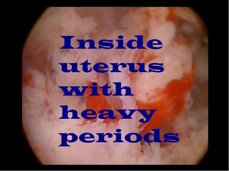
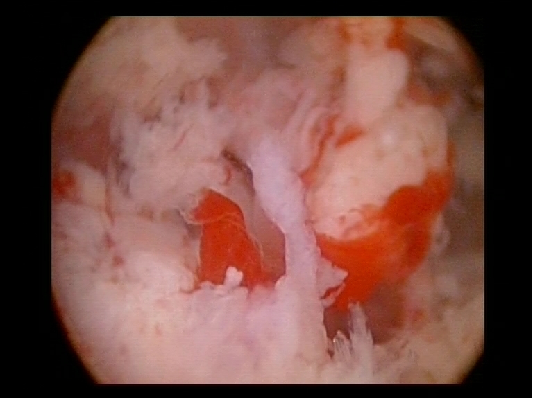
menorrhagia
|
|
|
|
endometrial hyperplasia with polyps
|
|
|
|
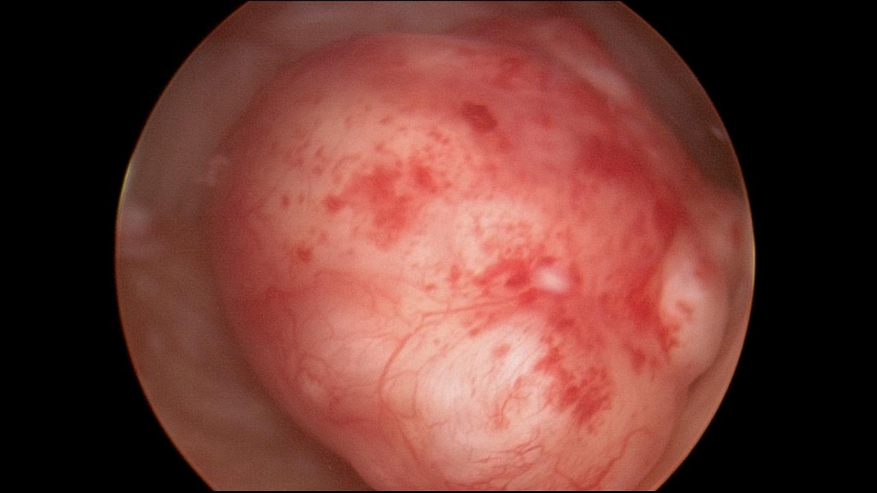
Submucus Fibroid
|
|
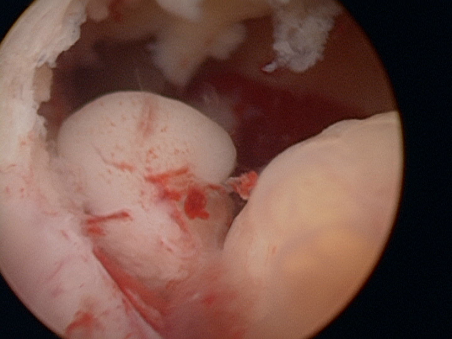
|
|
Endometrial Cancer 4
|
|