Laparoscopy is usually performed under general anesthesia; however it can be performed with other types of anesthesia that permit the patient to remain awake.
The typical pelvic laparoscopy involves a small 5mm-12mm incision in the belly button or lower abdomen. The abdominal cavity is filled with carbon dioxide. Carbon dioxide causes the abdomen to swell which lifts the abdominal wall away from the internal organs, so the doctor has more room to work.
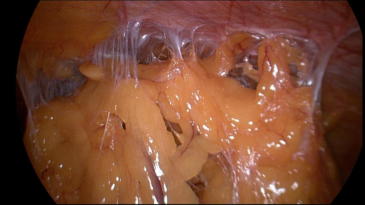
Next, a laparoscope (5-10mm fiber-optic rod with a light source and video camera) is inserted through the belly button incision. The video camera permits the surgeon to see inside the abdominal area on video monitors located in the operating room.
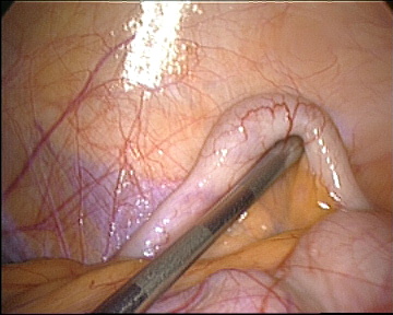
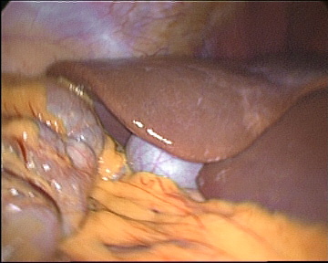
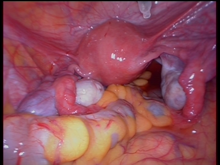
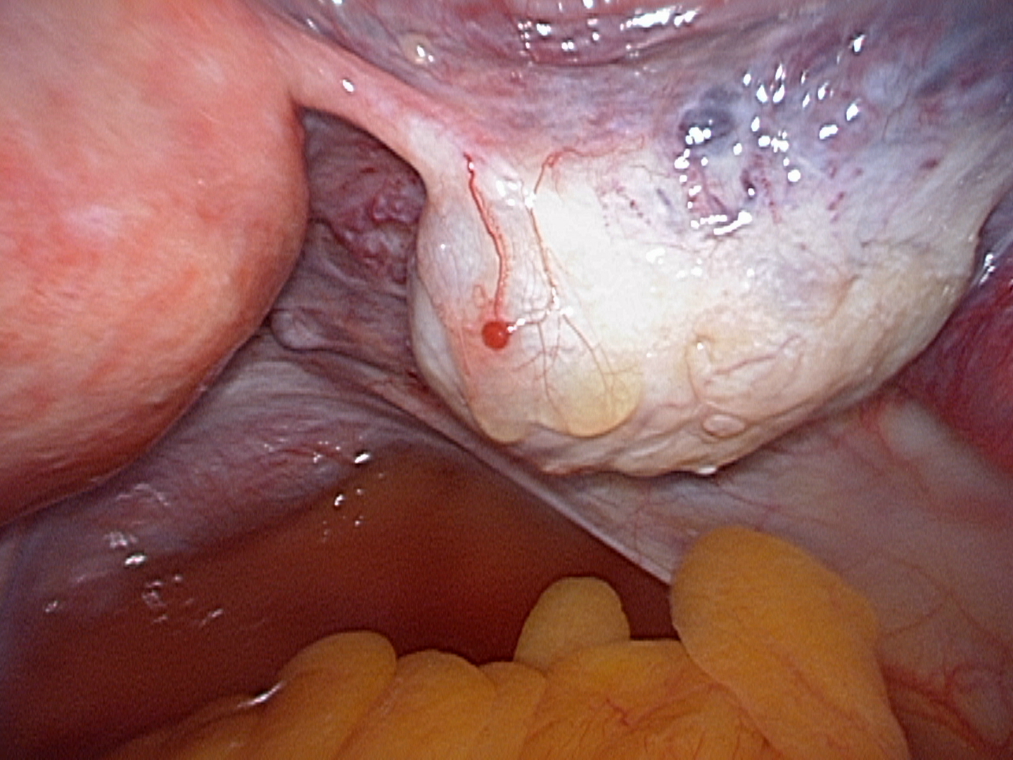
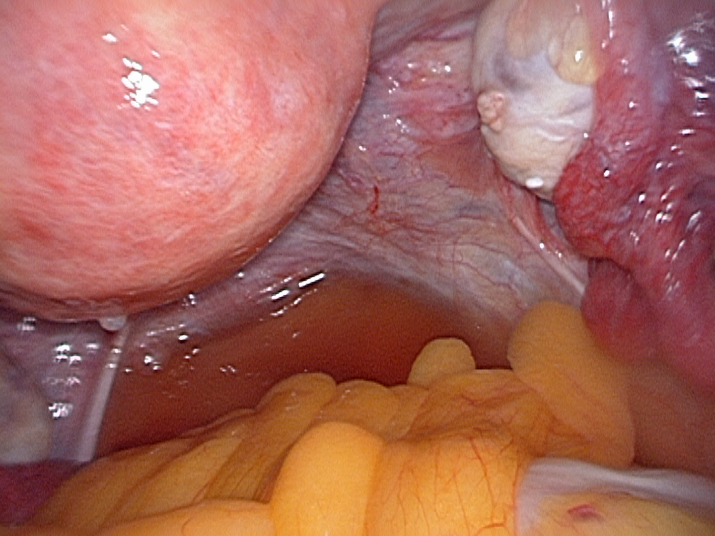
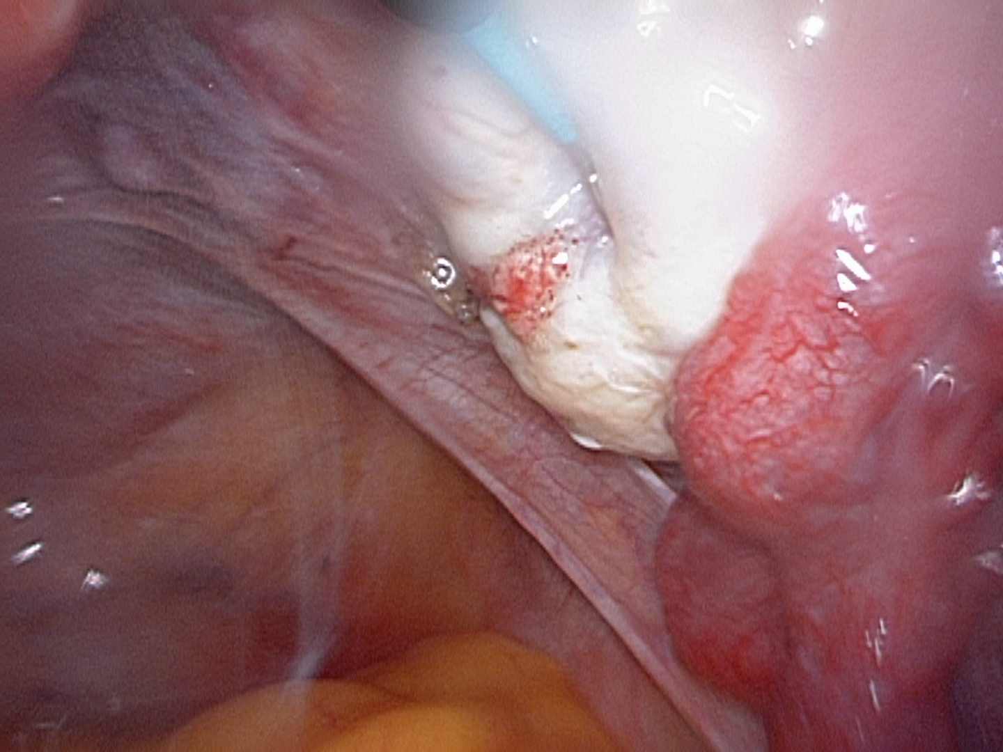
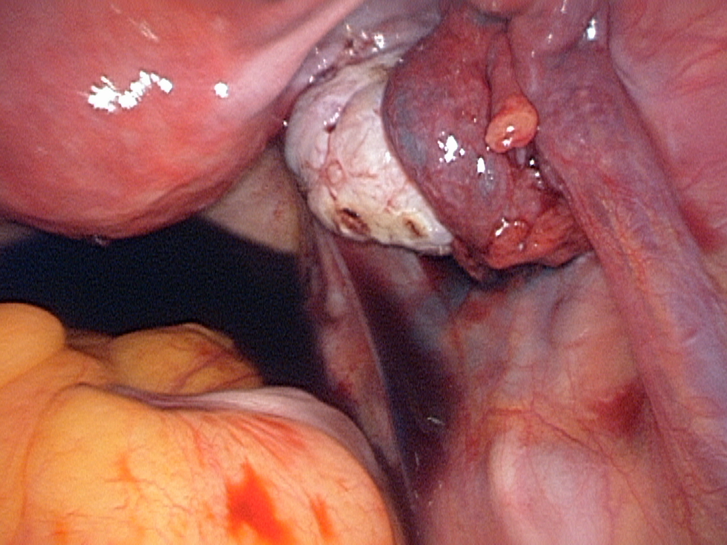
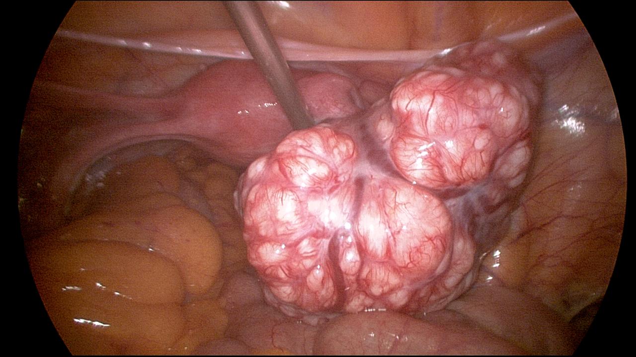
Depending on the reason for the laparoscopy, the doctor may perform surgery through the laparoscope by inserting various instruments into the laparoscope while using the video monitor as a guide. The video camera also allows the surgeon to take pictures of any problem areas he discovers.
