

The diagram illustrates anatomy of pelvic floor viewed from above. Ligaments of the pelvis support the vagina,cervix and pelvic organs. The ligaments include the Midurethral or Pubourethral ligament (PUL), the Arcus Tendineous (ATFP) and the Cardinal (CL) or Transverse Cervical ligaments and Uterosacral (USL) complex. The bladder rests on the Mid vaginal hammock (MVH). The uterus and cervix (CX) play a critical role in supporting the pelvic organs. Weakness or laxity of vaginal ligaments leads to prolapse and also prevents muscles from contracting effectively to open and close the urethra and anus. In the pelvis the muscles are not always attached directly to bone but transmit their forces indirectly via these ligaments.
Index
Incontinence
Prolapse
Fibroids
Period Problems
Ovarian Cysts
Endometriosis
Adhesions
Vaginal Discharge
Laparoscopy
Hysteroscopy
Cystoscopy
Pap Smear
Dysplasia
Colposcopy
LETZ
Hysterectomy
Vaginal Hysterectomy
Abdominal Hysterectomy
Oophorectomy
Ovarian Cystectomy
Dilatation Curettage
Myomectomy
Salpingectomy
Adhesiolysis
Endometrial Ablation
Novasure
Baloon Endometrial Ablation
Menopause
Prolift
Cystometrogram
Urodynamics
Videourethrocystmetrogram
Incontinence Slings
TVT
Safyre
Monarc
Sacrospinouscolpopexy
Hysterosalpingogram
Know Your Gynaecologist
Youssif Gynaecologist Melbourne
Contact
Glossary
Gynaecologist Melbourne Serag Youssif
doncaster-gynaecologist-obstetrician
Northpark Hospital
Bundoora Hospital
St Vincent Hospital
Freemasons Hospital
Epworth Eastern Hospital
Delivering Excellence and Specialised
Services in Safe Prolapse Surgery
Services in Safe Prolapse Surgery
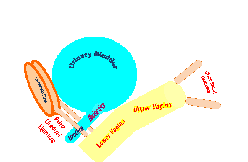
The cervix and uterus are held in the pelvis to side pelvic bones by two Cardinal ligaments [CL], to back bone the sacrum by two Uterosacral ligaments [ USL ] and to pubic bone in front by Pubocervical facia [PCF]. The urethra is held to back of pubic bone by midurethral ligament.
Illustration by Dr. Serag Youssif
Illustration by Dr. Serag Youssif
The lateral pelvic view shows how the vaginal tube is attached anteriorly to the symphysis pubis via the Pubourethral ligaments (PUL) and posteriorly to the sacrum via the uterosacral ligaments (USL).
Artwork by Dr Serag Youssif


